Rocks, Minerals and Soil Analyses Labs
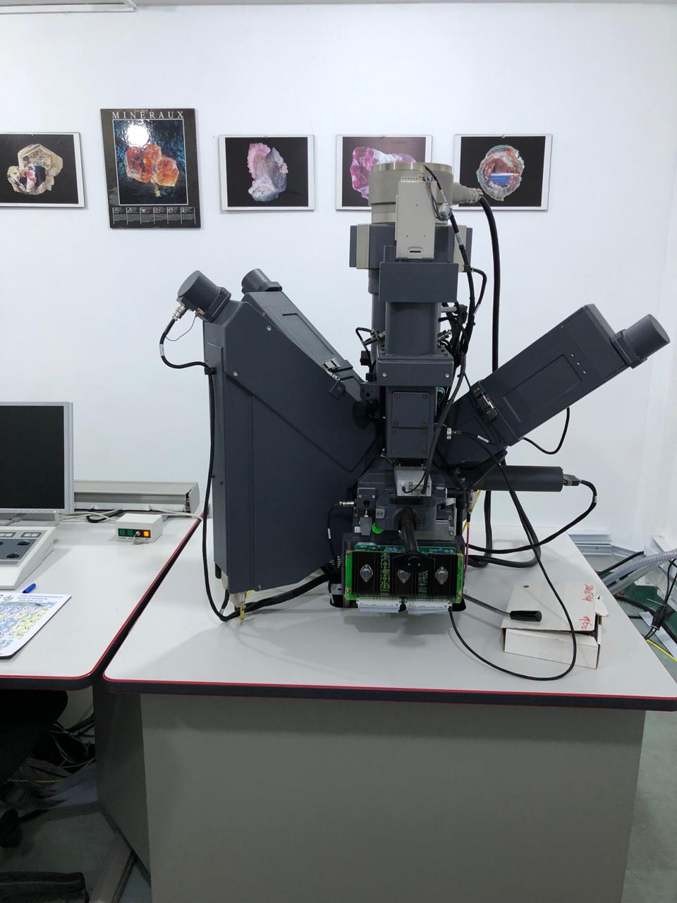
1. Electron Probe Micro Analysis (EPMA):
Electron Probe Micro Analysis (EPMA) is a sensitive non-destructive technology (< 100 ppm for most elements) used to analyze the chemistry of solid materials. It entails focusing an electron beam (usually with an energy of 5–30 keV) on a micro-volume (1-3 microns) of a specimen and then collecting and analyzing the X-ray radiation released by the different elemental groups using suitable crystal detectors. Because the X-rays' wavelengths are unique to the elements present in the sample, it is possible to establish the chemical makeup of the material. It is possible to identify any element with an atomic number of four or above. Uses in engineering and science include materials research, biology, electronics, geology, mechanical and chemical engineering, and electronics
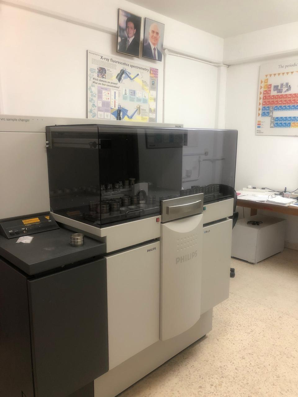
2. X-Ray Fluorescence:
X-ray Fluorescence (XRF) is a non-destructive analytical technique used for elemental analysis. In XRF analysis, a sample is irradiated with high-energy X-rays, which causes the atoms in the sample to emit characteristic X-ray fluorescence. By measuring the energy and intensity of these emitted X-rays, one can identify and quantify the elements present in the sample. XRF is widely employed in various fields, including geology, environmental science, archaeology, and materials science. Its versatility and rapid results make it a valuable tool for determining the elemental composition of a wide range of materials. It is particularly useful for quality control, alloy verification, and monitoring environmental pollutants. XRF analysis is appreciated for its speed and precision, making it an indispensable method for researchers and industries seeking to understand the elemental composition of their samples.
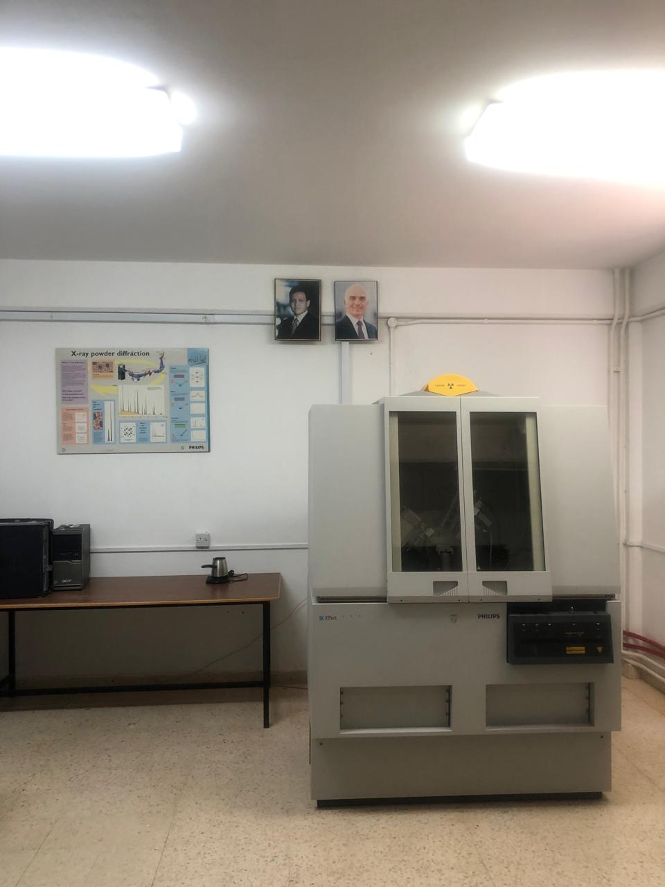
3. X-Ray Diffraction :
X-Ray Diffraction is used to determine the minerals and the chemical composition of
any crystalline powder or solid samples. It has a wide range of
applications in geology, material science, environmental science,
chemistry, and the pharmaceutical industry. Over 200,000 diffraction
patterns of known compounds have been compiled into a PDF database
which helps in the determination of the phases present in the sample by
modern computer programs through a quick compare of the diffraction
data of that sample to all of the patterns in the database. Analyzing
using the X-ray powder diffractometer is fast and accurate. A crusher
and a mill grinder are used for sample preparation.
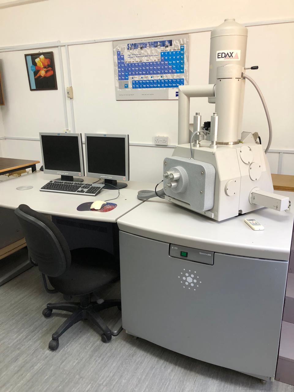
4. Scanning Electron Microscope (SEM):
Scanning Electron Microscope (SEM) is a type of electron microscope that produces images of a sample by scanning the surface with a focused beam of electrons. The electrons interact with atoms in the sample, producing various signals that contain information about the surface topography and composition of the sample.